
Benign Breast Conditions: The Role of the WHNP,” led by the author of this article, will be the focus of an Oct. 15 breakout session at the 24th Annual NPWH Premier Women’s Healthcare Conference, being held virtually from Oct. 13-16. Register here.
Nurse practitioners (NPs) working in women’s healthcare are often on the frontline of assessing, diagnosing, and managing benign breast disease. This article provides NPs with details on the systematic assessment, appropriate use of imaging studies and biopsy, and management of the benign breast mass.
Key words: benign breast mass, breast imaging, nonsurgical breast biopsy modalities
The most common benign breast conditions include benign breast masses or lesions, breast pain, nipple discharge, and breast skin changes. This article focuses on some of the most common palpable benign breast masses. These masses may be identified by the patient or by the nurse practitioner (NP) during clinical breast examination. It is critical to distinguish between benign and malignant masses and to know when referral to a breast specialist is indicated.
History and physical examination
When a patient presents with a complaint of finding a lump in the breast or other visible breast changes, a first step for the NP is to obtain a detailed history that includes breast cancer risk assessment. The history should include when the patient first noted the breast change, if the change is constant, intermittent, and/or cyclic, if further changes have occurred since initial detection, and if there are any associated symptoms (eg, pain, nipple discharge). Breast cancer risk assessment should include the patient’s personal menstrual, obstetric, and lactation history, personal history of any cancers, known inherited genetic mutations for breast cancer, breast biopsies, or breast density as determined by mammography, and family history of breast cancer or inherited genetic mutation for breast cancer.1
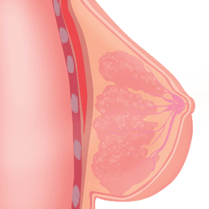
The NP should perform a thorough and systematic clinical breast examination. Intentional inspection of the exposed breasts and palpation of the bilateral breasts and axillae should be performed in both seated and supine positions. Masses should be assessed for size, texture, mobility, delineation, and tenderness. The NP should be alert for any other abnormal findings such as breast thickening, asymmetry, skin changes including erythema or peau d’orange appearance, retraction, and nipple discharge.1 Assessment of any nipple discharge should include determining color and consistency and if it is spontaneous or expressed only, unilateral/bilateral, and uniductal or multiduct. Breast examination findings should be documented in a systematic manner. Masses or skin changes should be described using the face of a clock to document position in the breast and distance in centimeters from the nipple.
Imaging studies
Breast imaging is usually necessary for a definitive diagnosis when there is a breast mass. This may include mammography with or without tomosynthesis, ultrasound, and/or breast magnetic resonance imaging (MRI). The National Comprehensive Cancer Network guidelines provide detailed algorithms for choosing appropriate breast imaging based on symptoms, clinical examination findings, and the patient’s age.2
Diagnostic mammography is used in women presenting with a palpable mass, focal mastodynia, or nipple discharge. This imaging modality is useful in detecting calcifications, masses, and architectural distortion. Tomosynthesis, or three-dimensional (3-D) mammography, is becoming increasingly utilized as it has been shown to increase detection of invasive cancers and decrease the rate of recalls for additional views.3
Breast ultrasound is useful for further evaluating palpable masses or changes identified on mammography. These images help to confirm if a mass is solid or cystic, which guides management decisions. Ultrasound is often the primary method of breast imaging in women younger than age 30 years with a palpable mass because breast density in this age group can significantly limit the interpretation of a mammogram. Based on sonographic characteristics, a mass can be determined to be benign, probably benign, or recommended to be biopsied.4
Breast MRI may be utilized for various indications and uses neovascularity of tumors and contrast enhancement for detection of breast cancer. It is utilized in breast cancer screening in high-risk populations; evaluating for occult breast cancers; determining the extent of disease in a known breast cancer; for further evaluation when imaging, clinical examination, and biopsy findings are discordant; and for evaluation of breast implant integrity. It is important to educate a patient on the high sensitivity and low specificity of MRI for breast cancer to prepare them for potential incidental findings and false positives that may lead to subsequent biopsy.5
The Breast Imaging, Reporting, and Data System (BI-RADS) is a standardized reporting system used by reading radiologists to provide management recommendations based on imaging findings. All dedicated breast imaging is assigned a BI-RADS category based on specific imaging characteristics and further classifies management and the likelihood of malignancy (Table).6 This lexicon provides management guidance regarding the lesion in question, whether a mass, calcifications, or architectural distortion. For any BI-RADS category 4 or 5 lesion, biopsy is indicated. Biopsy should be performed with core needle biopsy or fine-needle aspiration (FNA).2
| Table. Concordance between BI-RADS assessment categories and management recommendations6 | ||
|---|---|---|
| Assessment | Management | Likelihood of cancer |
| Category 0: Incomplete – need additional imaging evaluation and/or prior mammograms for comparison | Recall for additional imaging and/or comparisons with prior examination(s) | N/A |
| Category 1: Negative | Routine mammography screening | Essentially 0% likelihood of malignancy |
| Category 2: Benign | Routine mammography screening | Essentially 0% likelihood of malignancy |
| Category 3: Probably benign | Short-interval (6-month) follow-up or continued surveillance mammography | > 0% but _< 2% likelihood of malignancy |
| Category 4: Suspicious
Category 4A: Low suspicion for malignancy Category 4B: Moderate suspicion for malignancy Category 4C: High suspicion for malignancy |
Tissue diagnosis | > 2% but < 95% likelihood of malignancy
> 2% to _< 10% likelihood of malignancy > 10% to _< 50% likelihood of malignancy > 50% to < 95% likelihood of malignancy |
| Category 5: Highly suggestive of malignancy | Tissue diagnosis | _> 95% likelihood of malignancy |
| Category 6: Known biopsy-proven malignancy | Surgical excision when clinically appropriate | N/A |
Breast biopsy
Nonsurgical breast biopsy modalities for palpable masses include FNA, core needle biopsy, and vacuum-assisted biopsy. The choice of biopsy modality depends on mass characteristics and imaging findings. For masses that are not palpable, the same biopsy modalities may be used guided by ultrasound, stereotactic mammogram, or MRI.
FNA may be performed with or without local anesthetic, followed by aspiration with a 21- to 27-gauge needle and a 10- to 20-cc syringe. FNA is most often utilized for well-
circumscribed masses believed to be simple cysts. If the aspirated fluid is clear, it does not need cytologic evaluation. If the fluid is not clear or if the mass is solid, a specimen should be submitted for cytologic evaluation. Specimens obtained through FNA, however, frequently have insufficient cells for diagnostic cytology and high rates of false negatives.7
Core needle biopsy is the preferred nonsurgical method for solid masses because it provides sufficient tissue sampling for pathologic diagnosis. Core needle and vacuum-
assisted biopsies are performed by injecting local anesthetic and making a small incision in the skin to insert a 9- to 14-gauge needle for tissue sampling. The number of samples obtained varies and is determined based on the size and location as well as other factors such as mobility and consistency of the lesion. After a breast biopsy, a small tissue marker that is visible on mammography and MRI will be placed to mark the lesion for excision or for future follow-up on breast imaging. Tissue markers, or clips, are nonpalpable and are MRI compatible.
For any breast lesion identified on breast imaging and biopsy, clinical, radiologic, and pathologic concordance is essential.8 This will help guide decision making in the management of the lesion with either excision or observation. In certain instances, a lesion may be declared benign on core needle biopsy due to the limited tissue available for pathologic diagnosis. Breast imaging may demonstrate more suspicious features, however, such as mammographic spiculations or sonographic findings suggestive of a malignant process. Clinical exam may also demonstrate malignant features such as a firm fixed mass, skin changes, or lymphadenopathy. If clinical or radiologic features are worrisome for malignancy, repeat biopsy or excisional biopsy is imperative to rule out a cancer diagnosis.
Common benign breast masses
Breast cysts, or fluid-filled sacs within the breast tissue, are among the most commonly found breast masses and are almost always benign. Simple breast cysts are often identified in women in their 40s, but may occur at any time throughout the lifespan and are often hormonally fluctuant. These cysts may be identified as a palpable and sometimes tender mass or found on breast imaging or biopsy performed for another concern. Management of simple cysts can be symptom based, given that they usually resolve without treatment. If the patient complains of focal pain at the site of the cyst, if there are breast contour changes given size or location, or if the cyst is enlarging, the standard treatment is ultrasound-guided cyst aspiration, which also aids in confirming benign diagnosis.9 If cyst aspiration is performed, however, it is important to educate the patient that the cyst may re-accumulate and repeat aspiration may be necessary. In patients with cysts that continue to recur after aspiration, surgical removal can be considered.10
Galactoceles, another type of breast cyst, may be encountered in lactating women. These are milk-filled cysts within the breast that are typically caused by a clogged milk duct. Aspiration of the fluid can aid in diagnosis and provide relief of symptoms. Treatment with antibiotics should be provided in cases of infection. Galactoceles do not need to be surgically excised and will typically resolve on their own once the duct becomes unclogged.11 The patient may continue to breastfeed or pump.
Fibroadenomas are the most common of the benign solid breast masses, are often identified in adolescents and young adult women, and account for nearly 50% of all breast biopsies. These fibroepithelial lesions are typically solitary, well-
circumscribed, 1 to 2 cm in diameter, nontender, freely mobile, and firm or rubbery in consistency.9 Nipple discharge is not associated with fibroadenomas. Simple fibroadenomas confirmed by core needle biopsy can be managed conservatively with observation for growth or the development of other breast symptoms. If a fibroadenoma creates physical deformity, causes the patient distress, or demonstrates rapid growth, surgical excision may be indicated. Ultrasound-guided percutaneous excision or ultrasound-guided cryoablation are other treatment options.12
Phyllodes tumors, also fibroepithelial lesions, are less common than fibroadenomas, occur most often in women in their 40s and 50s, are generally larger, and show rapid growth. Phyllodes tumors should be managed by surgical resection with wide excision due to the risk of recurrence. If confirmed to be benign with clear margins, no further treatment is required.13 In about 5% of cases, phyllodes tumors can have malignant features and may require further oncologic treatment to reduce the risk of recurrence or metastasis.9
Intraductal papillomas may manifest as solitary lesions or can be found as multiple lesions in a patient with papillomatosis and are most common in the perimenopausal age group. Because these lesions obstruct and distend the involved lactiferous duct, intermittent and spontaneous nipple discharge is common. The discharge may involve single or multiple ducts and is typically watery, serous, or bloody. In fact, nipple discharge may be the patient’s only presenting complaint as the lesions are soft, small (2 to 4 mm in diameter), and usually nontender.9 Mammography and nonsurgical biopsy are indicated to confirm the diagnosis. A solitary intraductal papilloma without atypia should be managed on a case-by-case basis. Typically, a simple intraductal papilloma does not require excisional biopsy, with historical data demonstrating that there is a 5% to 20% chance that pathology would be upstaged to atypia or malignancy.8
If an intraductal papilloma is associated with atypical cells on biopsy, surgical excision is recommended due to risk of upstaging to a breast cancer identified on the surgical pathology. In patients with bloody nipple discharge, excision is typically recommended. Removal may also be considered for papillomas that are palpable, if the patient is symptomatic with non-bloody nipple discharge, or in consideration of other breast cancer risk factors.8
Two less common benign breast entities that may manifest as breast masses are pseudoangiomatous stromal hyperplasia (PASH) and hamartomas. PASH is most common in premenopausal women and may manifest as a palpable, mobile, nontender solid mass on clinical examination or be found incidentally on imaging.14 If identified incidentally on imaging and the patient is asymp-
tomatic, excision is not routinely recommended. If suspicious characteristics are apparent on imaging or during clinical examination, however, complete excision is important to confirm benign pathology without adjacent atypia or malignancy.15
Finally, hamartomas are a mixture of adipose, fibrous, and glandular tissue that typically present as a nontender, well-circumscribed solid mass or are identified on breast imaging. Management of hamartomas is also based on symptoms. In cases in which a hamartoma is confirmed via imaging and biopsy and the patient is asymptomatic, no further imaging follow-up or excision is needed.16 Neither PASH nor hamartoma is associated with an increased risk of breast cancer. 17
Practice implications
For patients with a benign breast mass, reassurance of the noncancerous nature of the finding is key. Education should involve a detailed discussion about implications of the lesion, symptom management, follow-up, and whether it has any impact on future risk for breast cancer. The patient should be referred to a breast specialist anytime there is not concordance of clinical breast examination, imaging, and nonsurgical biopsy findings that confirm a benign process.
In cases in which findings are concordant for a benign diagnosis and not bothersome to the patient, the NP is able to educate and manage the patient in the general outpatient clinic setting.
Stefani E. Yudasz is an advanced practice provider at the Parkview Cancer Institute in Fort Wayne, Indiana, and Instructor of Nursing at Vanderbilt University School of Nursing in Nashville, Tennessee. The author has no actual or potential conflicts of interest in relation to the contents of this article.
References
- Bickley LS. The breasts and axillae. In: Bates’ Guide to Physical Examination and History Taking, 12th ed. Philadelphia, PA: Lippincott Williams & Wilkins; 2016:419-448.
- National Comprehensive Cancer Network. Breast cancer screening and diagnosis. Version 1.2019. http://nccn.org/professionals/physician_gls/pdf/breast-screening.pdf.
- D’Orsi CJ, Getty DJ, Pickett RM, et al. Stereoscopic digital mammography: improved specificity and reduced rate of recall in a prospective clinical trial. Radiology. 2013;266(1):81-88.
- Stavros AT, Thickman D, Rapp CL, et al. Solid breast nodules: use of sonography to distinguish between benign and malignant lesions. Radiology. 1995;196(1):123-134.
- Baltzer PA, Benndorf M, Dietzel M, et al. False-positive findings at contrast-enhanced breast MRI: a BI-RADS descriptor study. AJR Am J Roentgenol. 2010,194:1658.
- D’Orsi CJ, Sickles EA, Mendelson EB, et al. ACR BI-RADS® Atlas, Breast Imaging Reporting and Data System. 5th ed. Reston, VA: American College of Radiology; 2013.
- Gordon PB. Image-directed fine needle aspiration biopsy in nonpalpable breast lesions. Clin Lab Med. 2005;25(4):655-678.
- American Society of Breast Surgeons. Consensus Guideline on Concordance Assessment of Image-Guided Breast Biopsies and Management of Borderline or High-Risk Lesions. November 2, 2016. https://www.breastsurgeons.org/docs/statements/Consensus-Guideline-on-Concordance-Assessment-of-Image-Guided-Breast-Biopsies.pdf.
- American College of Obstetricians and Gynecologists’ Committee on Practice Bulletins—Gynecology. Practice Bulletin No. 164: Diagnosis and management of benign breast disorders. Obstet Gynecol. 2016;127(6):e141-e156.
- American Cancer Society. Fibrosis and simple cysts in the breast. September 10, 2019. https://www.cancer.org/cancer/breast-cancer/non-cancerous-breast-conditions/fibrosis-and-simple-cysts-in-the-breast.html.
- Harris JR, Lippman ME, Morrow M, Osborne K. Diseases of the breast, 5th ed. Philadelphia, PA: Lippincott Williams & Wilkins; 2014.
- American Society of Breast Surgeons. Management of fibroadenomas of the breast. April 29, 2008. https://www.breastsurgeons.org/docs/statements/Management-of-Fibroadenomas.pdf.
- Jang JH, Choi MY, Lee SK, et al. Clinicopathologic risk factors for the local recurrence of phyllodes tumors of the breast. Ann Surg Oncol. 2012;19(8):2612-2617.
- Bowman E, Oprea G, Okoli J, et al. Pseudoangiomatous stromal hyperplasia (PASH) of the breast: a series of 24 patients. Breast J. 2012;18(3):242-247.
- American Society of Breast Surgeons. Choosing Wisely campaign: The American Society of Breast Surgeons releases new list of five things clinicians and patients should question. January 8, 2018. https://www.breastsurgeons.org/resources/choosing_wisely.
- Masciadri N, Ferranti C. Benign breast lesions: ultrasound. J Ultrasound. 2011;14(2):55-65.
- Degnim AC, Frost M, Radisky D, et al. Pseudoangiomatous stromal hyperplasia and breast cancer risk. Ann Surg Oncol. 2010,17(12): 3269-3277.